December 17, 2015
ZURICH, Switzerland, Dec. 15 (UPI) -- Researchers found a type of electromagnetic field can prolong survival in brain tumor patients who have already been treated with chemotherapy, according to a new study.
Tumor-treating fields -- low-intensity, intermediate-frequency alternating electric fields delivered with transducer arrays applied to a patient's shaved scalp -- have previously demonstrated an anti-tumor effect in background studies, leading researchers at the University of Zurich to test the treatment with glioblastoma patients.
Glioblastoma is a fast-growing tumor affecting the brain or nervous system that is difficult to treat, with most patients dying within a year or two of diagnosis. According to researchers, all attempts at improving outcomes for patients in large clinical trials have failed, leading them to test the combination treatment in a study published in the Journal of the American Medical Association.
Early trials by a company that markets a TTFields device, Novocure Ltd., showed promise, leading researchers to test it with patients, Dr. John Sampson, a researchers at Duke University, wrote in an editorial published in the Journal of the American Medical Association with the study.
Doctors at 83 medical centers in Europe, Canada, Israel, South Korea and the United States worked with researchers at the University of Zurich to recruit 695 glioblastoma patients, treating 466 with TTFields and the chemotherapy temozolomide and 229 with temozolomide alone. Patients who received TTFields were exposed to them for about 18 h per day using four transducer arrays placed on their scalp and Novocure's portable device. All of the participants received temozolomide for 5 days in each 28-day treatment cycle.
Ending the trial based on results from an interim analysis, median progression-free survival was 7.1 months in the group receiving TTFields, as opposed to four months in the group receiving only temozolomide. "Given the survival benefit reported in this study, it should now be a priority to understand the scientific basis for the efficacy of TTFields," Sampson wrote. "Achieving this may require the development of robust and widely available large animal models for glioblastoma, which do not currently exist."
December 04, 2015
 A review article about the area of magnetic particle hyperthermia just appeared in Applied Physics Review: http://dx.doi.org/10.1063/1.4935688. A predictable success of this review in the community can be inferred from its comprehensiveness.
Specifically, Gauvin Hemery and Dr Olivier Sandre (Univ. Bordeaux) were in charge of reviewing the chemical synthesis and coating requirements and methods of magnetic nanoparticles (within the view of good medical practice and scaled-up production to conduct medical assays); Dr Daniel Ortega (iMdea nanoscience, Madrid) described the existing physical models of magnetic hyperthermia (MH) and the current research to improve them; Dr Eneko Garaio and Pr Fernando Plazaola (UPV-EHU, Bilbao) listed the different types of devices (magnetic inductors, generators, resonant circuits…) to address the needs of standardization of preclinical assays (on rodents) in academic laboratories and industries, and ultimately for medical assays (on humans); Dr Francisco Teran (iMdea nanoscience, Madrid) focused on the modifications and biological fate of magnetic nanoparticles when injected to cells and tissues, which is mandatory to know to relate in intro and in vivo MH experiments; and finally, Dr Périgo (Univ. Luxembourg) was in charge of coordinating all the parts (writing the introduction/conclusion and of transitions, homogenizing notations, etc…), and last but not least, of managing the scientific discussion between specialists of different fields.
Several companies are involved in the MH area, among which we can cite: MagForce™ (Berlin, Germany), NanoTherics™ (Newcastle, UK, enterprise co-founded by a faculty of the University of Florida, Pr Jon Dobson), Resonant Circuits™ (London, UK spin-off company of University College London), nanoScale Biomagnetics™ (Zaragoza, Spain)… This study benefited from the collaborative framework of the European COST action TD1402 on "Multifunctional Nanoparticles for Magnetic Hyperthermia and Indirect Radiation Therapy" (RADIOMAG).
November 22, 2015
 Delivering cancer drugs with nanoparticles should reduce side effects by bringing the packaged drugs directly to tumors instead of distributing them freely throughout a patient’s body. But while this therapy works remarkably well to shrink tumors in some patients, nanomedicines can produce little to no effect in others. A therapy’s success may depend on a given tumor’s ability to uptake and retain these nanoparticles.
Now, researchers from Brigham and Women’s Hospital and Massachusetts General Hospital, led by Omid C. Farokhzad and Ralph Weissleder, report a technique to determine whether a particular patient has a high-uptake or low-uptake tumor (Sci. Transl. Med. 2015, DOI: 10.1126/scitranslmed.aac6522).
Nanoparticles accumulate selectively in tumors thanks to differences between blood vessels in healthy and cancerous tissue. Normal blood vessels, Farokhzad explains, “have very tight junctions with their neighboring cells to make the blood vessels like the piping in your home: They don’t leak.” In a cancerous growth, he continues, “tumor blood vessels are growing so fast that they don’t make those tight junctions,” allowing nanoparticles to leak out into the tumor.
But just as some leaky pipes can flood your basement and others leave only a small puddle underneath your sink blood vessels vary among tumors. Not every tumor has vessels leaky enough to deliver a nanoparticle payload large enough to muster an effective attack on the cancer cells. So Farokhzad, Weissleder, and colleagues sought a way to quantify the leakiness of blood vessels in a tumor and predict how well therapeutic nanoparticles could work for a particular patient.
The team injected rodents bearing human tumors with an FDA-approved anemia treatment called Feraheme, which consists of magnetic iron oxide nanoparticles coated with carboxymethyl dextran. They tagged the particles with fluorescent dyes so they could use high-resolution microscopic imaging to track the distribution of the drug and determine how much of it accumulated in tumor tissue.
They then repeated this experiment to track the accumulation of polymeric nanoparticles, which served as models for cancer nanomedicines. When the team compared each set of images pixel by pixel, they determined that the magnetic particles could predict the distribution of the anticancer ones with more than 95% accuracy. Therefore, the team concluded that if the magnetic particles accumulate in a tumor, it is very likely that anticancer ones will as well.
The researchers suggest that oncologists could use magnetic resonance imaging in the clinic to trace Feraheme iron oxide particles in a patient. This would allow doctors to decide if that person will benefit more from nanomedicine or standard formulations of a drug.
Feraheme is an FDA-approved drug with few side effects, so “it is conceivable that the approach could be rapidly translated,” says Nicolas Bertrand, a nanoparticle researcher at MIT, who was not involved in the study.
The study, Bertrand continues, is “among the first to introduce the concepts of personalized nanomedicine.”
October 15, 2015
 The new International Journal on Magnetic Particle Imaging (IJMPI) is a future platform for publishing high quality research articles on MPI. This journal has its origins in the International Workshop on Magnetic Particle Imaging (IWMPI), which is a unique annual meeting where the scientific MPI community discusses recent highlights of their research. The scope of the IJMPI ranges from imaging sequences and reconstruction over scanner instrumentation and particle developments to pre-clinical and clinical applications.
The designated Editor-in-Chief is Prof. Tobias Knopp. Journal articles will be published online with open access under a Creative Commons License. Please start with the submission of research papers within the scope of the IJMPI immediately in order to share ideas and experiences with a focussed audience. The link for submission of your MPI articles is http://journal.iwmpi.org.
To check out the editorial issue / article, see here.
September 06, 2015
 Although nanoparticles research is ongoing since more than 30 years, the development of methods and standard protocols required for their safety and efficacy testing for human use is still in development. Margarethe Hofmann and co-authors wrote a review that covers questions on toxicity, safety, risk and legal issues over the lifecycle of inorganic nanoparticles for medical applications.
The following topics were covered: (i) In vitro tests may give only a very first indication of possible toxicity as in the actual methods interactions at systemic level are mainly neglected; (ii) the science-driven and the regulation-driven approaches do not really fit for decisive strategies whether or not a nanoparticle should be further developed and may receive a kind of “safety label”. (iii) Cost and time of development are the limiting factors for the drug pipeline. Knowing which property of a nanoparticle makes it toxic it may be feasible to re-engineer the particle for higher safety (safety by design). For more details, see here.
August 21, 2015
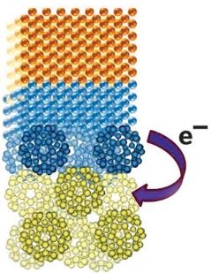 Only iron, cobalt, and nickel are permanently magnetic at room temperature, a property known as ferromagnetism that stems from how valence electrons are distributed and interact with each other within the metal. To develop or improve applications that use magnetism, such as computer memory and power generation, researchers would like to induce the property in other elements. Layering C60 on thin films of either copper or manganese successfully makes the metal layers ferromagnetic, reports a team led by Oscar Cespedes of the University of Leeds, in England (Nature 2015, DOI: 10.1038/nature14621).
The magnetization arises from electron transfer from the metal to C60, and it’s not limited to layering with buckyballs, Cespedes notes. Other materials with high electron affinity, as well as other metals, will also work. The effect depends on the thickness of the layers: The copper layer must be 2 to 3 nm thick, the manganese layer 2 to 15 nm thick, and the C60 layer 10 to 20 nm thick. The metal’s nonmagnetic bulk properties take over if the layers are too thick, and the magnetization drops off if the assemblies oxidize.
In the picture, a C60 nanolayer (green) induces magnetism in copper (orange) via electron transfer from copper to C60 at the interface between the two (blue).
July 27, 2015
 Immediately following the 250th ACS National Meeting in Boston, a one-day short course on surface modification will be presented. Professor John Texter has over 35 years experience in industrial small particle and coating technologies. He is Professor of Polymer and Coating Technology at Eastern Michigan University (since 2002).
The course is targeted at the advanced undergraduate and beginning graduate student level and will be useful to R&D chemists, materials scientists and engineers, and innovators in developing a toolbox of methods to use in optimizing formulations for advanced materials and applications.
Please see http://nanoparticles.org/courses/Boston2015Courses.htm
for details including course synopsis, outline, registration, and venue information.
July 24, 2015
 A. Paul Alivisatos and Alex Zettl of the University of California, Berkeley; Jungwon Park and David A. Weitz of Harvard University; and coworkers produced 3D images of angstrom-sized metal particles in solution at near-atomic resolution (Science 2015, DOI: 10.1126/science.aab1343).
The imaging technique used by the researchers, based on transmission electron microscopy (TEM), may enable scientists to monitor dynamics of individual particles within a colloid in their native state. Structural details gleaned from the imaging method may also lead to new uses for nanoparticles in catalysis, biological imaging, and other areas.
Drawing on a liquid-sampling technique the researchers reported in 2012 , they packaged a few droplets of colloidal platinum nanoparticles in a nanosized graphene bubble. Then they used a microscope equipped with a highly sensitive detector to zoom in on individual particles and record many short-exposure images.
Because the particles were constantly moving, the images were inherently low quality. But by applying a computational technique the researchers developed for this purpose, they were able to produce high-resolution 3-D reconstructions of randomly moving nonidentical particles.
This work is likely to make an impact in biological imaging, catalysis, and other fields, says Joerg R. Jinschek, a TEM specialist for microscope maker FEI who is based in the Netherlands. Jinschek explains that attaching a colloidal particle to a rigid surface—as has been done for imaging purposes in the past—can alter its structure. Because structure and other properties often depend on a particle’s state or environment, researchers want detailed information about nanoparticles in their natural states, Jinschek says. He adds that being able to image freely moving nanoparticles in liquids, as was done in the new study, will help scientists determine relationships between a nanoparticle’s structure and its function.
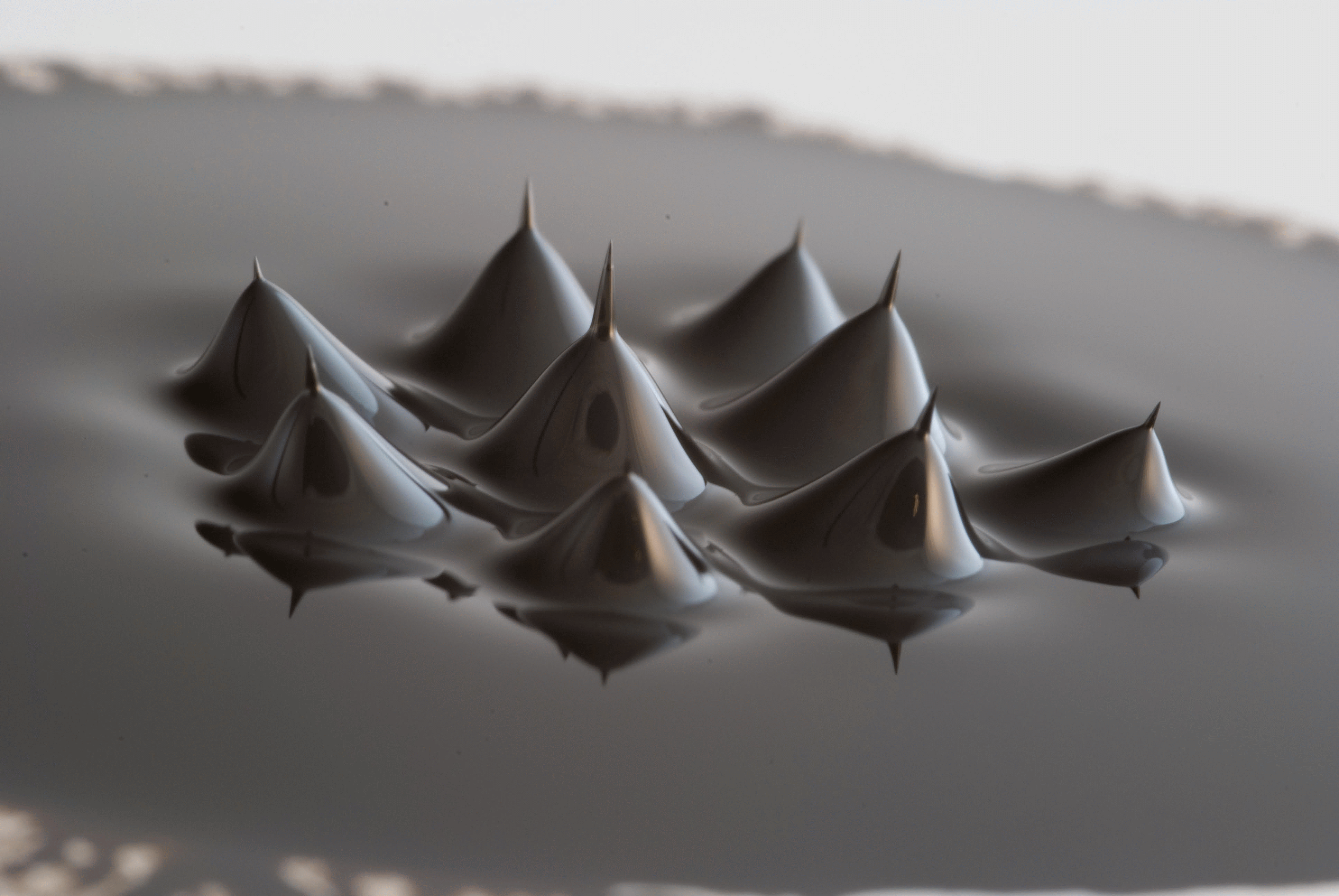
Beautiful ferrofluid, with a curious and striking 'peak-on-a-peak' effect. Submitted by Quentin Pankhurst.
Search this site with the power of
Last Modified: February 22, 2024 -
Magneticmicrosphere.com © 2025

