

Tiny Magnetic Sensors Might Open Door to Hand-Held Tests
November 13, 2016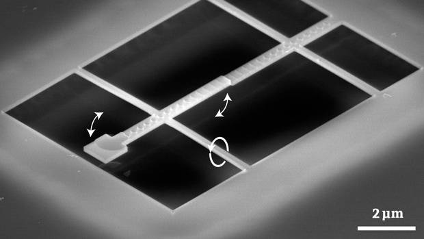 In 2008, Dr. Freeman and his team developed a tiny magnetic sensing device, called a torque magnetometer, on a piece of silicon chip that is smaller than the diameter of a strand of human hair. The device features a tiny spatula-shaped arm suspended on a narrow band of material that twists ever so slightly when the arm is pulled up or down by a magnetic field.
Prevention of Restenosis with Magnetically Targeted Endothelial Cells
October 06, 2016
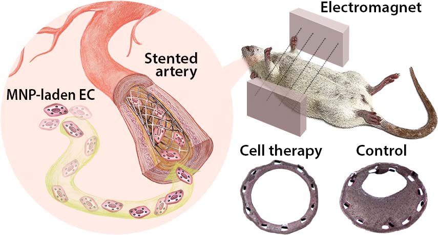 Boris Polyak at el. assessed the potential of magnetically mediated delivery of endothelial cells (ECs) to inhibit in-stent stenosis induced by mechanical injury in a rat carotid artery stent angioplasty model. ECs loaded with biodegradable superparamagnetic nanoparticles (MNPs) were administered at the distal end of the stented artery and localized to the stent using a brief exposure to a uniform magnetic field. After two months, magnetic localization of ECs demonstrated significant protection from stenosis at the distal part of the stent in the cell therapy group compared to both the proximal part of stent in the cell therapy group and the control (stented, nontreated) group: 1.7-fold (p < 0.001) less reduction in lumen diameter as measured by B-mode and color Doppler ultrasound, 2.3-fold (p < 0.001) less reduction in the ratios of peak systolic velocities as measured by pulsed wave Doppler ultrasound, and 2.1-fold (p < 0.001) attenuation of stenosis as determined through end point morphometric analysis.
Special Issue on Magneto-Plasmonics - Submissions Requested
August 07, 2016
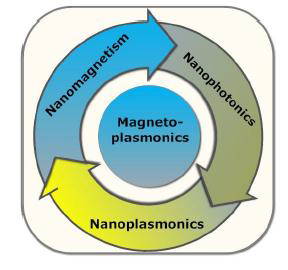 Magneto-plasmonics is a relatively new field that has great potential applications in biomedicine and biomedical technologies such as ultra-sensitive biosensing and bio-detection, bio-imaging, bio-therapy, drug-delivery, nano-imaging, to name a few. Deep understanding of various factors influencing magnetoplasmon properties is an important step in the effort to design new magnetic sensors and devices.
Although some progress on plasmonics has been achieved in the last few years, through combined simulation, modeling, experimental, and theoretical studies, there is still strong need to investigate new phenomena on magneto-plasmonics, in order to better tune and control magneto-optic properties, and to increase the sensitivity of the magnetic bio-sensor through modification of the optical radiation, magnetic field, and structure.
This new field merges the physics of nano-magnetics, where biological samples such as cells and DNA are made to interact with magnetic moments of a material in transverse direction, and nano-optics, where biological samples are made to interact with optical radiation in visible, infra-red, and telecommunication wavelength ranges. In a similar manner, it merges nano-plasmonics where biological samples are made to interact with surface plasmonic wave fields, also referred to as evanescent radiation fields.
Dr. Conrad Rizal from Baylor University's Department of Physics is the lead editor of this special issue. Deadline for paper submissions is November 1, 2016. Please check out more details here.
New Insight Into Magnetic Interactions During Magnetic Hyperthermia
July 09, 2016
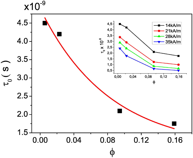 A new paper by Iacob, Kuncser, and Ladislau Vekas et al. carefully investigated, both theoretically and experimentally, the behavior of different concentrations of superparamagnetic nanoparticles in an alternating AC magnetic field ranging from 14-35 kA/m. They found that magnetic interactions, that increase with increasing volume fraction, can result in a decrease in SAR, whereas some authors claim that interactions can cause an increase in SAR.
See for yourself and read the paper here.
FDA Warns from Use of Anemia Drug Feraheme (Ferumoxytol)
June 13, 2016
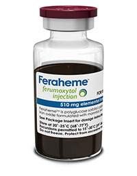 The U.S. Food and Drug Administration (FDA) is strengthening an existing warning that serious, potentially fatal allergic reactions can occur with the anemia drug Feraheme (ferumoxytol). We have changed the prescribing instructions and approved a Boxed Warning, FDA’s strongest type of warning, regarding these serious risks. Also added is a new Contraindication, a strong recommendation against use of Feraheme in patients who have had an allergic reaction to any intravenous (IV) iron replacement product. Health care professionals should follow the new recommendations in the drug label.
Check out this pamphlet here to read about it.
The effects were very serious, as you can learn from the last paragraph of the FDA warning: Since the approval of Feraheme on June 30, 2009, cases of serious hypersensitivity reactions, including death, have occurred. A search of the FDA Adverse Event Reporting System database identified 79 cases of anaphylactic reactions associated with Feraheme administration, reported from the time of approval to June 30, 2014. Of the 79 cases, 18 were fatal, despite immediate medical intervention and emergency resuscitation attempts. The 79 patients ranged in age from 19 to 96 years. Nearly half of all cases reported that the anaphylactic reactions occurred with the first dose of Feraheme. Approximately 75 percent (60/79) of the cases reported that the reaction began during the infusion or within 5 minutes after administration completion. Frequently reported symptoms included cardiac arrest, hypotension, dyspnea, nausea, vomiting, and flushing. Of the 79 cases, 43 percent (34/79) of the patients had a medical history of drug allergy, and 24 percent had a history of multiple drug allergies.
For more information, check out this website: http://www.fda.gov/Drugs/DrugSafety/ucm440138.htm
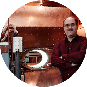
Obituary of Founding JMMM Editor Arthur J. Freeman
June 06, 2016 It is with a heavy heart that we must inform you of the death of JMMM's Founding Editor, Arthur J. Freeman, on June 7, 2016. He will be sorely missed for his scholarship, mentorship and friendship. Professor Freeman launched the Journal of Magnetism and Magnetic Materials in 1975 with North Holland (Elsevier) as publisher. The inaugural Editorial Board of the Journal read like a who's who in the %uFB01eld. Even so, launching a new journal is no easy task. It requires synergy between Editor and Publisher. Prior to this Professor Freeman had a false start with his International Journal of Magnetism, which was only published between 1971 and 1974 by a different publisher.
Will the MPI Inventors Get the European Inventor Award 2017 ?
May 04, 2016
 All of you probably know Bernhard Gleich, Jürgen Weizenecker and team who invented the Magnetic Particle Imaging (MPI) technique. We now have a chance of helping them to win the European Inventor Award 2017 in the industry section. Please go to
http://www.epo.org/learning-events/european-inventor/popular-prize.html
and vote for them. Would be great if we can help them to win this prestigious award!
And by the way, the video that they made about MPI and the possibilities of this technique in the future is very impressive, well worth the 6 minutes it takes to watch it!
Smart Shoe to Reduce Foot Ulcers
April 15, 2016
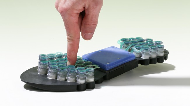 EPFL researchers together with the University Hospital in Geneva have developed a shoe sole with valves that electronically control the pressure applied to the arch of the foot, aiming at preventing foot ulcers commonly caused by diabetes. The sole has around 50 small electromagnetic valves filled with magnetorheological material. The viscosity of the material, which is made up of suspended iron microparticles, can be controlled by applying a magnetic field. The particles react immediately and align themselves with the field, causing the material to change from liquid to solid state in a fraction of a second. The system should not only help the wounds heal quickly but also prevent the onset of new ulcers. Every year, 250'000 diabetics have a leg amputated in Europe alone, mainly because of foot ulcers.
For more information, check out our Archives.
September 2017
Search this site with the power of