

Smartphone Takes Fluorescent Nanoparticle Pictures
October 02, 2013
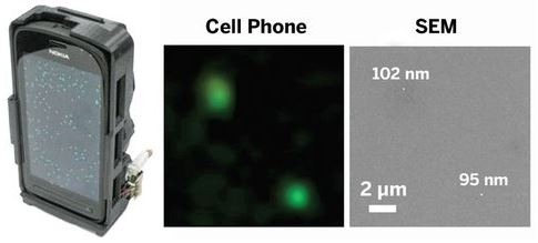 By attaching a small, inexpensive gadget to the back of a smartphone, scientists have created a sensitive handheld fluorescence microscope. The attachment allows the phone’s camera to take pictures of fluorescently labeled nanoparticles and virus particles, making the phone a potentially useful portable diagnostic tool for health care workers in developing countries ( ACS Nano 2013, DOI: 10.1021/nn4037706 ).
The device, designed by Aydogan Ozcan of UCLA and colleagues, consists of a laser diode to excite fluorescent dyes and a compact system of lenses and filters that remove background noise created by the laser light. The team used the microscope to detect human cytomegalovirus particles, which are between 150 and 300 nm in diameter, and polystyrene beads, which at about 100 nm in diameter were the smallest objects that could be detected. According to Ozcan, the mini microscope is the first portable, cell-phone-based imaging system sensitive enough to resolve individual nanoparticles and viruses. Ozcan started a company called Holomic to commercialize the device, and his group has created smartphone apps for data analysis.
Successful Meeting - Frontiers in BioMagnetic Particles III
August 29, 2013The Frontiers in BioMagnetic Particles III meeting was held in the idyllic location of Telluride CO, with participants from 9 different countries. A wide range of high quality invited and contributed talks were given on subjects including monodispersed nanoparticle fabrication, MRI and MPI imaging, drug delivery, magnetically modulated energy delivery (MagMED), and magnetically actuated materials. There was also a career development panel with participants from large industry and startup companies, government labs and agencies (NIST, Sandia, and NCI), and academia. Every session included a panel discussion on challenges and opportunities, with all participants in the session serving as panelists. Poster sessions were held outside with delicious food and drinks catered by the Telluride Sceience Research Center (TSRC) staff. The event provided excellent networking opportunities; social activities during and around the meeting included hiking, biking, gold panning, and ballooning. Overall the meeting was a great success!
To checkout the program, see here. And if you have questions about it or want to get on their mailing list, then please contact Thompson Mefford and Jeffrey Anker who organized the meeting flawlessly in one of the most scenic places! Their next meeting will take place in 2015. And now a few pictures from the meeting.

Varying Ferromagnetism in a Single Material
August 15, 2013Researchers at the Paul Scherrer Institute have managed to grow a material that is simultaneously ferromagnetic and antiferromagnetic. They grew layers of LuMnO3 on a crystal and found that close to the crystal the material exhibited ferromagnetism, but became antiferromagnetic farther away. The material's structure adapts itself to that of the underlying crystal, which creates this gradient. While varying ferromagnetism has previously been created by layering materials, this is the first time the feat was achieved in a single material. This has implications for the de-sign of compact digital storage media.
http://swissinnovation.org/news/web/2013/04-130712-65
South Korea Testing 'Electric Road' That Charges Buses as They Travel
August 13, 2013
 A South Korean city has begun testing an "electrified road" that allows electric public buses to recharge their batteries from buried cables as they travel. The Korea Advanced Institute of Science and Technology (KAIST), which developed the system, said it would be tested over the next four months on a 24-kilometre route in the southern city of Gumi.
Pick-up equipment underneath the bus, or Online Electric Vehicle (OLEV), sucks up power through non-contact magnetic charging from strips buried under the road surface. It then distributes the power either to drive the vehicle or for battery storage As a result it requires a battery only one-fifth the size of conventional electric vehicles. The system also eliminates the need for overhead wires used to power conventional trams or trolley buses. The technology does not come cheap, with each OLEV costing around $630,000.
"The technology is readily available but the question is how to bring down the cost," said Park Jong-Han, manager of the company that produced the OLEV prototypes. "Once the cost goes down, I believe more cities will be interested in commercialising the new transport network," Park told AFP. The system has already been partially trialled on a much smaller scale at an amusement park and on the KAIST campus.
Electrifying the road does not require major construction work, as the recharging stations only have to be buried along 10-15 per cent of the route at places such as bus stops.
Better Measurement of Single Cell Temperature
August 05, 2013
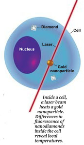 Nanoscale thermometry has untold possible uses for biology, from basic research on how heat flows in living systems, hyperthermia research in magnetic treatments, to controlling gene expression with temperature. Researchers use a number of techniques for biological thermal sensing, including Raman spectroscopy and detection of fluorescing proteins. But all have problems, such as low sensitivity or the inability to make highly localized measurements. Now, a group led by physics professor Mikhail D. Lukin and chemistry professor Hongkun Park at Harvard University have harnessed a common defect in diamonds to develop an improved approach for nanometer-scale thermometry in biological systems ( Nature 2013, DOI: 10.1038/nature12373 ). In some diamonds, two adjacent carbons are replaced by a nitrogen and an empty spot called a nitrogen vacancy center. Minuscule temperature changes strain the lattice of such diamonds, which affects the quantum spin properties of the local defect and modifies its fluorescence properties. These changes can then be detected.
The group used microwave pulses to manipulate a diamond-lattice defect’s spin states,
and from resulting changes in fluorescence, researchers determined corresponding
temperature variations. The method, they found, is capable of detecting temperature
changes as small as 1.8 mK in areas as small as 200 nm across. The team then tested the thermometer in a living human cell, an embryonic fibroblast. They inserted nanodiamonds and gold nanoparticles into the cell. Laser light heated the gold nanoparticles, and the group monitored temperature gradients throughout the cell by observing changes in the nanodiamonds’ fluorescence. “I like this technique very much,” says Xinwei Wang , a mechanical engineering professor at Iowa State University, noting that in addition to being very sensitive, the technique’s spatial resolution is comparable or superior to that of widely used Raman techniques.
“This kind of sensitivity is extremely important when diagnosing thermal responses and studying chemical reactions in biosystems at the cellular level,” Wang says. Konstantin V. Sokolov, a physics professor at the University of Texas, Houston, calls the work “a precious solution” to the problem of measuring temperatures in biological systems on the nanometer scale. With improvement, the team says the technique may make it possible to observe real-time biological activity with subcell resolution.
The 13th Ferrofluid Workshop is Coming Up !
July 23, 2013
 This year's and now already 13th Ferrofluid Workshop in Benediktbeuern, Germany will take place from September 25-27, 2013.
Check out the details here:
http://www.mfd.mw.tu-dresden.de/ffworkshop/
Tannic Acid and Fe(III) Form Special Coating on Almost Any Substrate
July 19, 2013
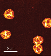 A team led by Frank Caruso at the University of Melbourne has developed a simple one-pot recipe for a coating made of only Fe(III) and tannic acid, a polyphenol found in wood that is perhaps best known for improving the flavor of wood-casket-aged red wine. The coating is unusually versatile. It can cover all manner of nano- and microscopic objects, including
gold nanoparticles, calcium carbonate and silicon dioxide particles, and bacteria, regardless of whether the object to be coated is positively charged, negatively charged, or neutral ( Science 2013, DOI:10.1126/ science.1237265). The discovery is patentpending.
Since both Fe(III) and tannic acid are generally regarded as safe by regulators and are already used in food and biomedical applications, the coating could find uses right away in areas as diverse as drug delivery and corrosion protection, comments Christopher W.
Bielawski , a chemist at the University of Texas, Austin.
“This is going to make a big impact mainly because it is so simple,” he adds. “Many will wish that they had thought of it first.”
“The coating’s pH sensitivity is the really exciting aspect,” comments Phillip B. Messersmith , a biomedical engineer at Northwestern University. The coating forms above pH 6, but in more acidic environments it falls apart, revealing or releasing the contents of objects within it. One could envision using the coating as a way to deliver a drug to a cell’s acidic lysosome or endosome and then having the contents released in the organelle’s low-pH environment, he says. Researchers use “tannic acid” to describe a family of molecules that contain a central glucose with one to five polygalloyl groups of varying lengths that emanate from the sugar base. Every Fe(III) atom can coordinate three pairs of hydroxyl groups found on tannic acid and thus can complex up to three different tannic acid molecules. Meanwhile, the abundance of hydroxyl groups in tannic acid means that each tannic acid molecule can complex up to a dozen or so iron atoms. The result is a cross-linked coating that is about 10 nm thick, Caruso says.
The technique seems like it could be easily expanded, Bielawski says. “There are a lot of other polyphenols out there besides tannic acid, so there’s considerable potential for changing the surface chemistries of the coating and creating new applications.”
Improve Energy Conversion with the Help of Magnetic Nanoparticles
July 15, 2013
 AN OBSTACLE to developing low-cost energy conversion devices is understanding why small differences in materials can strongly affect performance. Figuring out the relationship between the structure and function of an electrode material, for example, is especially challenging for nanoparticle aggregates.
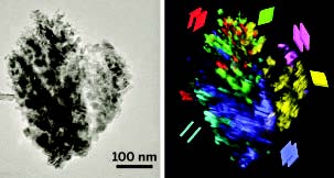
Researchers have now developed an analytical procedure to scrutinize material structure and function with nanometer resolution across micrometer distances in such aggregates. The technique enables them to design a better electrode for solar energy conversion. The group used transmission electron microscopy (TEM) to image the spatial distribution and orientation of nanocrystals within aggregates.
Then, by using a conducting atomic force microscopy method, they correlated the TEM information with the pathways that electrons follow as they move through the material. They used the combo method to probe electron transport in various nanoparticle-based iron oxide electrodes. The electron-transport proficiency of this cheap material makes it useful for generating hydrogen via light-driven water splitting. Although the electrodes they studied were similar, some worked well, while others did not.
The key finding of the team, which includes Scott C. Warren of the University of North Carolina, Chapel Hill, is that the relative orientation of adjacent nanoparticles greatly affects charge transport. A small orientation mismatch is alright, but a large mismatch results in electrical barriers that block current flow between adjacent grains. The upshot is that by identifying “winning” crystal orientations and tailoring the preparation method to favor them throughout the electrode, the group made a device that achieves a record-setting photocurrent for this class of materials ( Nat. Mater. 2013, DOI: 10.1038/nmat3684).
“These results represent an important step forward in developing nanostructured materials for next-generation energy conversion devices,” says solar-fuel specialist Roel van de Krol of Technical University of Berlin. Boston College chemist Dunwei Wang adds that the study helps explain why some nanostructures function better than others. “This work will be of value to photoelectrochemistry studies in general,” he adds. — MITCH JACOBY, C&EN
For more information, check out our Archives.
September 2017
Search this site with the power of