

Stabilization of the Surfaces of Magnetite
December 20, 2014
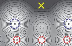 Accurate structures of iron oxide surfaces are important for understanding their role in catalysis, and, for oxides such as magnetite, applications in magnetism and spin physics. The accepted low-energy electron diffraction (LEED) structure for the surface of magnetite, in which the bulk surface termination undergoes an undulating distortion, has a relatively poor agreement with experiment. Bliem et al. show that the LEED structure is much more accurately described by a structure that includes subsurface cation vacancies and occupation of interstitial sites (see the Perspective by Chambers). Such cation redistribution occurs in many metal oxides and may play a role in their surface structures.
Check it out at Science 5 December 2014:Vol. 346 no. 6214 pp. 1215-1218, DOI:10.1126/science.1260556
Handling of Magnetic Beads in Microfluidics
December 15, 2014
 Spinomix SA, a Swiss technology platform company announced the approval of two US patents while three others are pending approval. The two US Patents are number US 8,585,279 and US 8,870,446 relating to the manipulation and mixing of magnetic particles in microfluidics environments.Spinomix provides innovative sample processing solutions to the life sciences sector. The company’s unique MagPhase technology enables homogenous handling of magnetic beads in microfluidics based systems, thus enhancing bioassay efficiency and in a further step, allowing for sample processing automation. MagPhase technology is currently applied in nucleic acid purification, a market estimated to represent over one billion dollars. The vision of the company is to expand the use of microfluidic cartridges into additional areas, such as protein purification or cell isolation.
To read more, check out Spinomix website.
Smart Application of Magnetic Fields Allows for Deep Central Focusing of Magnetic Nanoparticles
December 15, 2014
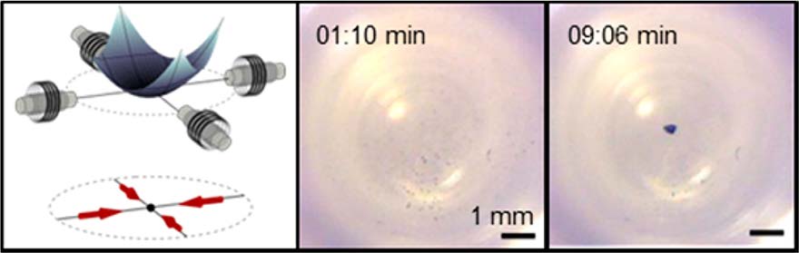 Recent efforts between the University of Maryland (UMD) and Bethesda-based Weinberg Medical Physics LLC (WMP) have led to a new technique to magnetically deliver drug carrying particles to hard-to-reach targets. The method has the potential to transform the way deep-tissue tumors and other diseases are treated. UMD Fischell Department of Bioengineering (BioE) alumnus Dr. Aleksandar Nacev and BioE and Institute for Systems Research Professor Benjamin Shapiro have teamed up with WMP to exploit fast pulsed magnetic fields to focus nano-therapeutic magnetic particles to deep targets.
Pulsed magnetic fields allowed the team to reverse the usual behavior of magnetic nano-particles. Instead of a magnet attracting the particles, they showed that an initial magnetic pulse can orient the rod shaped particles without pulling them, and then a subsequent pulse can push the particles before the particles can reorient. By repeating the pulses in sequence, the particles were focused to locations deep between the electromagnets.
To find out the details for yourself, check out the Nano Letters paper which is available online at http://dx.doi.org/10.1021/nl503654t with a video showing the magnetic focusing at http://ter.ps/magnetic.
Webinars by Malvern
December 02, 2014
 Many of us own size determination equipment from Malvern, for example DLS based instruments. Or since the addition of NanoSight to Malvern a year ago, one of their instruments. But you might not have seen their informative webinars on the latest technologies, applications, and research yet. They are now all available on Malvern's website, and you can watch the recorded version at your leisure here.
For examples, recent titles are:
The roles of nanoparticles in therapeutic protein aggregation pathways
Powerful protein SEC made simple!
Speed your way through your protein formulation screening by automating your measurements
Miltenyi Celebrates 25th Anniversary
November 26, 2014
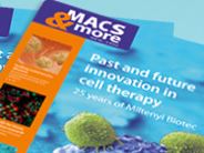 Wow, Miltenyi is already 25 years old - how the time flies. They fittingly celebrate their achievements by publishing a MACS anniversary issue where they highlight how helpful magnetic particles are in today’s most promising approaches to cellular therapies, involving regulatory T cells, NK cells, stem cells, neural cells, and CAR-expressing T cells. The field of immunotherapy is becoming more and more important, as being able to regulate the behaviour of these cells will help to treat cancer, autoimmune diseases.
Miltenyi Biotec revolutionized cell processing for both basic research and clinical application. Their techniques help to unleash xenograft technology, which is a leap forward in cancer research. In their anniversary issue of MACS, you can check on two fold-out pages the milestones that got Miltenyi Biotec from their first product, the superparamagnetic biotin Microbeads, different columns and the MACS separator which allowed for magnetic isolation of cells, to today, with now fully automated systems for cell isolation, flow cytometry, cell soring and molecular analysis. A remarkable story. Check out their anniversary issue !
Google X Reveals Nano Pill To Seek Out Cancerous Cells
November 24, 2014
 Detecting cancer could be as easy as popping a pill in the near future. Google’s head of life sciences, Andrew Conrad, took to the stage at the Wall Street Journal Digital conference to reveal that the tech giant’s secretive Google[x] lab has been working on a wearable device that couples with nanotechnology to detect disease within the body.
“We’re passionate about switching from reactive to proactive and we’re trying to provide the tools that make that feasible,” explained Conrad. This is a third project in a series of health initiatives for Google[x]. The team has already developed a smart contact lens that detects glucose levels for diabetics and utensils that help manage hand tremors in Parkinson’s patients.
The plan is to test whether tiny particles coated “magnetized” with antibodies can catch disease in its nascent stages. The tiny particles are essentially programmed to spread throughout the body via pill and then latch on to the abnormal cells. The wearable device then “calls” the nanoparticles back to ask them what’s going on with the body and to find out if the person who swallowed the pill has cancer or other diseases. For more, click here.
Granules of Iron Oxide Chitosan Particles Remove Arsenic, Microbes, and Other Contaminants in Simple-to-Operate System
November 10, 2014
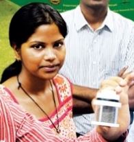 Groundwater in the Indian state of West Bengal naturally contains arsenic, causing ailments including skin diseases and cancer. Thanks to nanotechnology, thousands of people there have gained access to arsenic-free water since 2013, with the installation of treatment tanks using porous granules developed by a team at the Indian Institute of Technology (IIT), Madras, led by chemistry professor Thalappil Pradeep. The technology has received government support for field-testing as an option for low-cost, point-of-use water treatment.
The granules are nanocomposites made from ferric oxyhydroxide and a biopolymer, chitosan. Iron oxides remove arsenic ions from water by adsorption. The team boosted their metal oxyhydroxide’s activity by reducing the particle size to nanoscale, thereby increasing the surface-to-volume ratio, and anchoring the material within a network of chitosan. With this structure, which resembles sand and is made at room temperature, embedded particles don’t leach into water, and the captured arsenic stays put. What goes on “in the atomic scale is not completely understood,” Pradeep says, but that has not stopped the material’s real-world use.
At the Ambattur industrial estate, in a suburb of the Indian city of Chennai, a facility makes about 36 kg of the ferric oxyhydroxide-chitosan nanocomposite per day. Production at the plant—run by InnoNano Research, a start-up founded by the IIT Madras team—is enabling field trials in West Bengal. For more information, check DOI: 10.1073/pnas.1220222110.
Report from the Benediktbeuern Colloquium 2014
October 21, 2014
 From September 29th to October 1st 2014, the 2nd Colloquium of the DFG Priority Program 1681: Field controlled particle matrix interactions: synthesis multi-scale modelling and application of magnetic- hybrid materials was held in the Bavarian cloister Benediktbeuern. This colloquium is part of a special program of the German Research Foundation (DFG) (i.e., DFG Priority Program 1681) that started in January 2014 and is focused on novel magnetic hybrid materials research. The research ranges from production to technical and medical applications and includes modelling of field dependent interaction with different matrices. The work benefits from the cross-specialization collaboration of chemists, physicist, engineers, biologists, and medics.
Nearly 9 months after the start of the program, more than 60 scientists from each of the 27 projects in the program presented their most recent research findings in scientific talks and posters. The scientific reports presented during the colloquium showed very promising results. The highlight of the three-day meeting was a hiking tour in the mountains that culminated in scientific presentations being given in an alpine hut (without any projection equipment). For the selected presenters, it was an honor to speak in this unusual setting as its technical limitations require extra clarity in the communication of results.
The next colloquium will take place at the end of September 2015 at which time the first 2-year funding period will be coming to a close and groups will be looking to apply for more funding on the basis of their results.
Link to SPP description: http://www.mfd.mw.tu-dresden.de/spp1681/index.php/willkommen
For more information, check out our Archives.
September 2017
Search this site with the power of